Shunts
Most shunts currently available respond to differences in pressure between the ventricle cavity that they drain and the cavity to which the shunt drains to (so-called pressure differential valve shunts). There are many standard pressure differential valve shunts currently available, with only slight differences between them. There is no evidence that any one company's design is better that another and, in fact, some companies carry more than one design. The most recent technological improvement in shunt design has been the introduction of systems where the shunt's valve and reservoir comes with an integral distal catheter, thus eliminating the need to attach a distal catheter to the valve/reservoir at the time of surgery. This improvement is expected to drastically reduce the incidence of distal catheter disconnection and consequent shunt malfunction. With the availability of this newer design, most pediatric neurosurgeons have abandoned the used of pieced-together systems when implanting a new system or completely replacing an old system.
Commercially available shunts are categorized as low, medium or high pressure, depending on their response to the pressure differential between upper and lower ends of the shunt. Theoretically, a low-pressure shunt drains a pressure of 0 mm of water, a medium-pressure one at 60 mm, and a high-pressure one at 120 mm. However, although these figures may be roughly correct when the patient is recumbent, there is a complete change in the upright position, as the system then resembles a siphon tube being used to drain an auto's gasoline tank. The result is that the pressures within the head drop into the negative range as CSF is sucked out of the head by siphoning, and overdrainage of the ventricles can occur. This is true regardless of which type of simple pressure differential valve is utilized. There is no physiologic correlation between the type of valve, the intracranial pressure and the outcome of surgery. Nor is there at this time any convincing evidence that one pressure setting is better than another. Our preference has been for low-pressure systems in a newly diagnosed neonate, but other surgeons have been equally successful with other systems.
Overdrainage of the ventricles can result in collapse of the brain away from the inner surface of the skull, with a resultant risk of bleeding with compression of the brain. In the long term, overdrainage can result in headaches of a very debilitating degree. Consequently, several design modifications of the pressure differential valve have been developed. First, an anti-siphoning device was developed to be added to the standard shunt just below the shunt's reservoir. This device is responsive to atmospheric pressure as transmitted through the skin. When the pressure within the fluid column of the shunt drops to below atmospheric pressure (as is the case with siphoning), atmospheric pressure transmitted through the skin pushes a diaphragm down to close the fluid passageway and block further flow of CSF through the system until such time as the pressure within the ventricles climbs back to above atmospheric pressure. When this occurs, the diaphragm is pushed back open. A variation on this is the Pudenz-Schulte Delta valve shunt. This shunt has a valve on it very similar to the anti-siphon device in that it resists drainage through the system when pressures within the system drop into the negative range. It has a theoretical advantage over the anti-siphon valve in that it does not need to perceive atmospheric pressure to work, and thus encasement of the valve by scar tissue does not cause its malfunction as opposed to the anti-siphon valve where this has been reported. Also available is a valve that closes when a person is standing and opens when he or she is lying down (the horizontal-vertical valve). It is typically used in shunts running from the lower spinal canal into the abdominal cavity (so-called lumboperitoneal shunts) in individuals with communicating hydrocephalus. Another system, NMT’s Orbis valve, offers a variable resistance to flow as a function of variations in pressure within the ventricles. As pressure increases within the ventricles, the resistance to flow increases until pressures reach an abnormally high range where the valve drops its resistance to flow to zero to allow decompression of the dangerously high state. This design is meant to avoid overdrainage due to transient, normal rises in pressure within the head, such as happen when an individual coughs or sneezes. Recently, a multicenter study took place in North America comparing the rates of failure in the Orbis-Sigma and Delta valve shunts to rates seen in standard pressure differential valve shunts. There was no difference in failure rates, and no particular shunt was proven better than any of the others.
The newest design in shunt valves is the Codman Hakim programmable shunt. This shunt has a pressure differential valve whose resistance can be altered using a magnet field transmitted through the skin. Thus, the valve settings can be changed during a routine office visit, avoiding any surgery. Reports from Europe and North America have stated that some patients have benefited from having this shunt. Clearly, siphoning occurs with this system, so it is unclear to what degree the feature of valve adjustment will be of benefit. One potential use for this system is in the treatment of normal pressure hydrocephalus (NPH), a condition seen in adults. Here a low-pressure shunt is desired, but the implantation of such a device can lead to acute overdrainage and the risk of bleeding over the surface of the brain. This system allows for the gradual introduction of a low-pressure shunt by starting out at a high resistance setting and slowly moving downward. This shunt has also been used in individuals suffering from chronic headaches and a functioning shunt. Recently, Codman has introduced the Hakim programmable valve with a siphon-guard valve. This system has both a programmable pressure differential valve and an anti-siphon valve distal to it. It is too early to comment on when this type of system is optimally used.
Typically, the upper end of the shunt is placed to the brain's fluid chambers using surface landmarks about the head as guides. The catheter enters the head via holes drilled through the skull (burr holes). These holes are behind the hair line, either at the top of the head, behind the ear or in the back of the head. Flow of CSF out of the tubing confirms that the tube's tip is inside the brain’s fluid chambers. Occasionally, more sophisticated guidance of the tube is required. Small endoscopes can be inserted into the catheter and used to visually guide the shunt's tip to a point within the brain's ventricles (fluid chambers), thus optimizing positioning. Image guidance systems, which use CT or MR scans obtained pre-operatively to help the surgeon visualize where within the brain the catheter is, are also available when precise positioning is required.
Several body cavities are available for distal drainage of a shunt. When shunts were first introduced almost 40 years ago, a one-way valve drained spinal fluid directly into the right atrium of the heart via the jugular vein (ventriculoatrial shunt). Vascular shunts functioned very well, but they were prone to multiple problems, including early and late infection, as well as rare, potentially fatal heart failure due to blockage of blood vessels within the lungs by particles of blood clot flaking off the shunt's catheter tip. The use of the heart has been largely abandoned as an initial choice because of these problems, but it remains a viable second option when infection or surgery has rendered the abdominal cavity unaccommodating of the distal shunt catheter. The chest cavity is another cavity that can be used as a backup to the abdominal cavity (ventriculopleural shunt). The catheter is placed inside the rib cage between its inner lining and the outer lining of the lungs. Occasionally, this cavity cannot resorb the CSF rapidly, and the lung becomes compressed by the excess CSF, resulting in difficulty in breathing. The catheter must be moved to a different cavity is such cases. Rarely, the catheter can rest on the diaphragm (the muscle at the base of the lungs used for breathing), causing irritation and hiccups. Finally, recent reports from Europe describe placing the distal end of the shunt into the major blood sinuses (e.g., the sagittal sinus) draining blood from the brain.
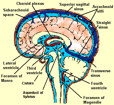
The rationale for this is to duplicate the normal outflow resistance experienced by cerebrospinal fluid as it is reabsorbed into the sinus via arachnoidal villi. This work is based on work originally done by Hash in 1979 and Wen in 1982. An Egyptian physician, El-Shafei, has also had extensive experience with this type of system.
The peritoneal cavity is now the most common distal site for shunt placement (ventriculoperitoneal shunt). It is a large cavity, more than capable of handling any amount of CSF delivered by the shunt in all but the most unusual cases. The rhythmic contractions of the intestinal organs tend to move the tip of the shunt catheter around the abdomen, thus minimizing the changes of it becoming sequestered in scar tissue and subsequently blocking. As the child grows, changes in the length of the torso are accommodated by tubing being pulled out of the abdominal cavity. There has been an evolution in thought about how much catheter should be placed in the abdominal cavity of infants and children. Whereas a few years ago only 8 or 12 inches of tubing was placed into the cavity, it is now accepted that a neonate may have 36 or more inches in the peritoneal cavity (i.e., enough tubing to accommodate adult stature without the tube's end being pulled out of the abdominal cavity. There have been no associated complications, and mandatory lengthening is no longer necessary.
Chronic Headaches and Shunts
A significant number of shunt-dependent children and adults are plagued by chronic, often disabling headaches. These are usually not associated with other symptoms of increased intracranial pressure, and neuro-diagnostic studies usually do not reveal evidence of a shunt malfunction. In most cases the CAT or MRI scan shows small ventricles. The condition of having small ventricles on CAT or MRI scan coupled with chronic headache complaint has been termed slit ventricle syndrome.
Small ventricles are a desirable result of shunting, but they can sometimes cause problems. There are a number of therapeutic options. In the first place, it is essential that a CAT scan be carried out while the patient is symptomatic. This is because an intermittent shunt malfunction may masquerade as slit ventricle syndrome, with the fluid spaces enlarging only at the time of malfunction. Scans done when the individual is asymptomatic will show the ventricles to be unchanged in comparison to scans done previously when the patient was asymptomatic. Other causes for headaches, such as sinusitis, are searched for on the scan.
Recent evidence has shown that shunt-dependent children with chronic headache are often suffering from a migraine equivalent. They do not experience classic migraine, but may for some reason be vulnerable to similar variations of in blood volume within the brain. It has been found that anti-migraine therapy (Inderal, etc.) has a dramatic result in many such cases. Therefore, we now recommend that this regime be employed with all children experiencing chronic headache in the absence of obvious shunt malfunction. Intracranial pressure monitoring and surgery can also be considered when other avenues have been exhausted.
Once it has been documented that the ventricles are unchanged during symptomatic periods and pharmacologic treatment has failed, the next step is to determine what is happening to the pressures within the head (intracranial pressure) during periods of headache. Intracranial pressure monitoring is exceedingly useful in evaluating chronic headache complaints in shunt-dependent children and adults. We have found that this symptom may be produced by higher-than-normal pressure (hypertension) or lower-than-normal pressure (hypotension), or it may be unrelated to the shunt's function. Appropriate treatment is obviously dependent on identifying the true cause of the symptoms, and intracranial pressure should be determined in all such cases.
Intracranial hypotension may occur many years after insertion of the shunt as a result of axial growth and consequent increased siphoning through the shunt; it is treated by increasing the resistance of the system or placing an anti-siphon device in the system. Intracranial hypertension can be more problematic to deal with. First, it is confirmed that a CAT or MRI scan has been obtained while the individual was maximally symptomatic to rule out an intermittent shunt malfunction. If this has been done, consideration is given to changing the shunt's valve if it is not a low-resistance system. Finally, if headaches persist and pressure monitoring shows episodes of severe rise in intracranial pressure with resulting headache, either surgical expansion of the skull or establishing of bone widows under the temple's muscles can be done to allow underlying expansion of the brain to dissipate transient periods of increased pressure. In cases where pressure monitoring fails to document changes in intracranial pressure during periods of headache, the child or adult is referred to a headache clinic with the reassurance that there is no problem with the shunt's function.
Long-Term Care of Shunts
Periodic visits to the neurosurgeon are desirable to monitor the shunt’s function over time. Generally, it is obvious when a shunt is malfunctioning. Signs of an overt failure would include recurring, intensifying headache and/or irritability, lethargy, nausea/vomiting with loss of appetite, and loss of the ability to look upward. Subtle symptoms can also suggest a problem with the shunt. These would include an unexplained decline in work or school performance, pain or redness along the shunt tract, or fever of an unexplained origin. An examination will frequently uncover signs of increased pressure within the head when a shunt malfunctions.
Plain X-rays will demonstrate rupture or disconnection of the shunt's catheters, and CAT or MRI scans can demonstrate interval change in ventricular size. When a CAT or MRI scan is performed, it is imperative to compare it with a scan taken when the child was feeling well and the shunt was working properly (see example). It is not unusual for scans thought initially to be normal to in fact show an interval enlargement of the fluid spaces due to malfunctioning of the shunt. This can be appreciated only when current scans taken when the individual was symptomatic are compared with scans done when he or she was feeling well. Blood tests can be used to see if there are signs of an inflammatory process within the body, as would occur with an infection, and CSF can be withdrawn from the shunt's reservoir to look for signs of infection within the CSF.
A properly functioning shunt does not require manipulation of the pump. If the system is draining sluggishly, a decision is sometimes made to have the family speed it up with the pump. But this is a temporary expedient; revision due to a shunt element's malfunction is almost inevitable. In addition, the pumping of the shunt to evaluate its function can be misleading. There is often little correlation between function and what is noted on depressing the pump. If a child is asymptomatic, there is no need to manipulate the apparatus, and this may in fact cause problems with drainage.
Shunts commonly function for many years without problems of any sort. And so, of course, parents often inquire whether the child "still needs the shunt." The answer is almost always yes. The overwhelming majority of children will remain shunt-dependent all their lives. Possible exceptions to this rule are those with spina bifida; for unknown reasons approximately 20 percent of them develop arrested hydrocephalus.
In a few patients, serial abdominal X-rays will reveal that the shunt has become distracted from the peritoneal cavity. A decision must then be made as to whether or not the tube is to be lengthened. If the CAT scan discloses a small ventricular system, there is no question that the shunt is functional, and it is reasonable to lengthen it before the child becomes symptomatic. Arrested hydrocephalus is invariably associated with moderately to markedly enlarged fluid spaces, so that normal or small ventricles are always indicative of shunt dependency.
If a child is diagnosed as having arrested hydrocephalus, it is incumbent on the physician to ensure that no damage to the nervous system occurs through incorrect diagnosis. These patients should have annual neuropsychological testing, looking for a subtle decline in higher cortical function. Any suspicion of an evolving deficit mandates an immediate shunt revision.
Many years ago, when shunts were made of relatively brittle materials, there was a tendency to be conservative about encouraging sports or other vigorous activity. However, it has become clear that present-day shunts are not easily fractured, and limiting sports in children who are physically able to engage in them is a significant emotional burden. We encourage our patients to participate in soccer, tumbling, gymnastics or anything else they may choose.
Children with shunts are, of course, vulnerable to all the usual illnesses of childhood. When these are accompanied by high fever, it is sometimes suggested that the shunt be tapped to exclude the possibility of infection of the shunt. However, it should be kept in mind that 95 percent of shunt infections occur within three to five days of surgery, so this is a most unlikely etiology.
Fever persisting longer than expected in the absence of an obvious explanation may occasionally call for shunt aspiration in spite of this unlikelihood. If so, it is essential that the sample be obtained from the pump rather than with a spinal tap. The latter will frequently test negative even when there is an active shunt infection.
It should also be pointed out that shunt-dependent children are very prone to headaches during febrile illness or vigorous activity, and this should be a source of concern only when the headaches persist and/or worsen.
The presence of a shunt in a female is not a contraindication to her becoming pregnant, and, if this is her desire, she should be supported in it. When a woman with a shunt becomes pregnant, she can experience an increase in the frequency and intensity of headaches due to changes in her intravascular volume and, as a result, her cerebral blood flow. Because this state can be difficult to differentiate from a shunt malfunction, it is wise to obtain an MRI scan at the beginning of pregnancy to be used as a baseline for comparison should symptoms of increased intracranial pressure arise.